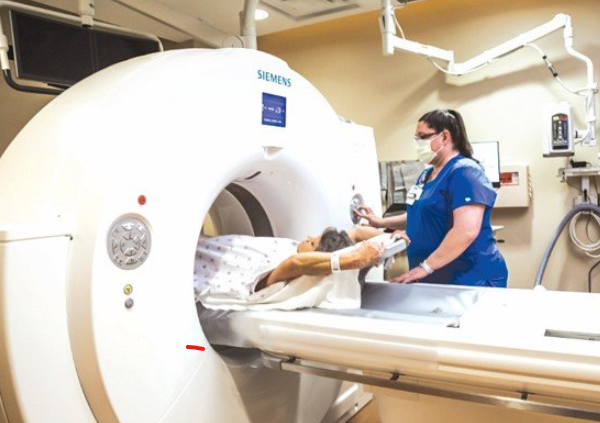
As lung cancer cases continue to rise in Kenya and around the world, experts are urging citizens to adopt advanced imaging techniques such as chest CT scans and Positron Emission Tomography (PET) scans for screening. These methods can facilitate early detection of the disease and assess the effectiveness of ongoing treatments. Early detection is vital for improving survival rates, as it enables timely interventions and more precise treatment planning.
“Despite advances in imaging technology, diagnosing lung cancer remains challenging due to the potential for false positives and negatives,” says Dr Solomon Mutua, a clinical oncologist at
Nairobi West Hospital.
“Benign lung nodules can sometimes resemble malignant tumors on a chest CT scan, leading to misdiagnosis.”
He added that “lung cancer can also be hidden within inflammatory changes or scarring in the lungs”.
“That is why use of advanced imaging techniques is recommended to enhance detection and accuracy.”
Although a chest X-ray is the quickest, most cost-effective, and widely used initial imaging test for the lungs, it has limitations compared to advanced techniques.
While a chest X-ray can detect some lung tumors, it may overlook small, early-stage tumors and is less effective at evaluating the spread of cancer. In contrast, a chest CT scan provides detailed images by capturing multiple pictures of the lungs and chest, which are then compiled into a comprehensive view. This advanced imaging technique allows for the detection of very small nodules and is particularly beneficial for diagnosing lung cancer at its most treatable stage.


